

General and Obstetric Ultrasound

Overview | Obstetric Ultrasound | Preparation | What to Expect | Results and Follow-Up

What Is Ultrasound?

The Women’s Imaging Center performs general ultrasound and obstetric ultrasound exams in Mount Vernon, Washington. Ultrasound, also called sonography, uses sound waves to create real-time images of internal organs, soft tissues, blood flow, and pregnancy-related anatomy.

Ultrasound does not use radiation. Because of this, it is often used as an initial imaging exam when appropriate, depending on the area being evaluated and the reason for the study.
ultrasound

Common Ultrasound Exams

General ultrasound may be used to evaluate many areas of the body, including the abdomen, pelvis, thyroid, kidneys, blood vessels, and soft tissues. Your provider may order an ultrasound to help evaluate symptoms such as pain, swelling, abnormal lab results, a palpable area, or another clinical concern.

Common ultrasound exams may include:

  • Abdominal ultrasound
  • Carotid ultrasound
  • Pelvic ultrasound
  • Thyroid ultrasound
  • Vascular ultrasound
  • Venous ultrasound

Because ultrasound provides real-time images, it can help assess structure, movement, and blood flow depending on the type of exam being performed.

Obstetric Ultrasound

Obstetric ultrasound, sometimes called an OB sonogram, may be used to evaluate fetal anatomy, fetal position, gestational age, and other pregnancy-related questions your provider may have.

The exam is noninvasive and generally well tolerated. Preparation may vary depending on the stage of pregnancy and the type of exam being performed. Our team will provide instructions when your appointment is coordinated.

Preparing for Ultrasound

Preparation instructions vary by exam type. Some abdominal ultrasound exams may require fasting for several hours before the appointment. Some pelvic or kidney ultrasound exams may require a full bladder.

If preparation is needed, our team will provide instructions before your visit. Following preparation instructions helps improve image quality and supports a more complete exam.

What to Expect During an Ultrasound

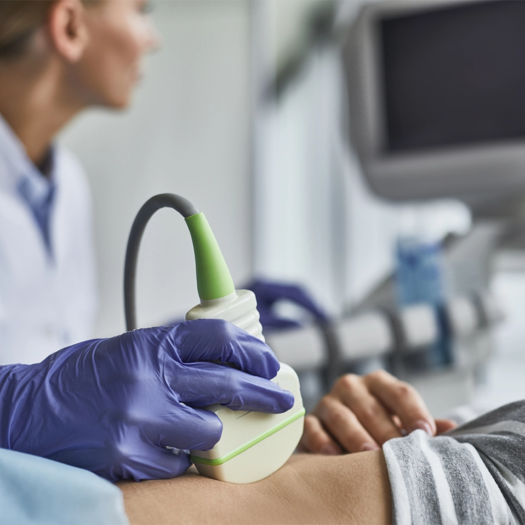
During an ultrasound exam, gel is applied to the skin over the area being evaluated. A small handheld device called a transducer is moved over the area to create images. You may feel light pressure during the exam, especially if the area being evaluated is tender or if a full bladder is required for the study.

The technologist will obtain the images needed for the radiologist to review. The exam length may vary depending on the type of ultrasound and the area being evaluated.

Results and Follow-Up

A radiologist will review the ultrasound images and send a report to your referring provider. Your provider will review the findings and next steps with you. Depending on the reason for the exam, recommendations may include routine care, additional imaging, follow-up, or referral to another provider.

To request an appointment or ask questions about ultrasound preparation, please request an appointment online or call (360) 428-7275. A member of our team will follow up to help coordinate your appointment.