Our Services

Breast Cancer Screening

Breast cancer is the most common cancer in women regardless of race or ethnicity. One in 8 women in the United States will develop invasive breast cancer. 85% of breast cancers occur in women with no family history. Studies have shown that screening mammograms save lives, reducing mortality from breast cancer by 20-40%.

Recommendations for when to start screening and how often to screen vary by different medical societies.

  • The American College of Radiology (ACR) recommends annual screening mammograms at age 40 for women at average risk for developing breast cancer.
  • The American Cancer Society recommends annual screening mammography for women at age 45 and possibly earlier at age 40 based on a discussion with their doctor.
  • It is recommended that patients with strong family histories of breast cancer consult with their doctors for risk assessment as earlier screening is often recommended.
SYSIMG-006

3D Mammography

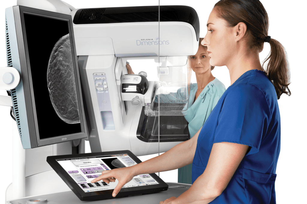
Tomosynthesis, also known as 3D mammography, is an advanced form of mammography which takes multiple images of the breast from different angles. This allows doctors to evaluate the breast tissue layer by layer. Instead of viewing all of the complexities of your breast tissue in a flat image, as with conventional 2D mammography, fine details are more visible and no longer hidden by the tissue above or below. The low radiation dose is comparable to that of a 2D mammogram.

3D mammography is more accurate and has been shown in studies to have a higher cancer detection rate as well as lower recall and false-positive rates compared to 2D mammography. In addition, 3D mammography is better at detecting smaller cancers before they have spread to lymph nodes.

Breast Ultrasound

Breast ultrasound utilizes sound waves to obtain images of your breast tissue. It is used as a complementary test with mammography to evaluate specific regions of the breast. It is generally not as sensitive for detecting breast cancer as mammography but can be used to characterize lesions seen on mammogram or provide an additional look at focal areas of pain or lumps palpated by the patient.

Since there is no radiation associated with ultrasound exams, they are also used to evaluate the breast in young patients with lumps or pain.

Breast MRI

Breast MRI utilizes magnetic fields and radio waves to generate detailed images of your breast tissue. It is very sensitive and is utilized as a complementary test with mammography.. Breast MRI is often performed to evaluate the extent of disease when a patient has been diagnosed with breast cancer. It is also used to characterize lesions seen on mammography or ultrasound which are indeterminate. In patients at high risk for breast cancer including those with strong family histories, breast MRI may be offered as an additional screening test in addition to mammography.

Breast Biopsy

Once a lesion is found in the breast that cannot be characterized as benign, a breast biopsy is required to obtain tissue samples for analysis by a pathologist to determine whether it is a form of cancer or benign finding. Because most of these lesions are small at the time of detection, imaging guidance is necessary to ensure proper sampling of the correct region. Biopsies may be performed under ultrasound guidance, mammographic guidance (stereotactic biopsy), or MRI guidance. These procedures are performed at the Women's Imaging Center by our team of breast imaging radiologists.

General Ultrasound

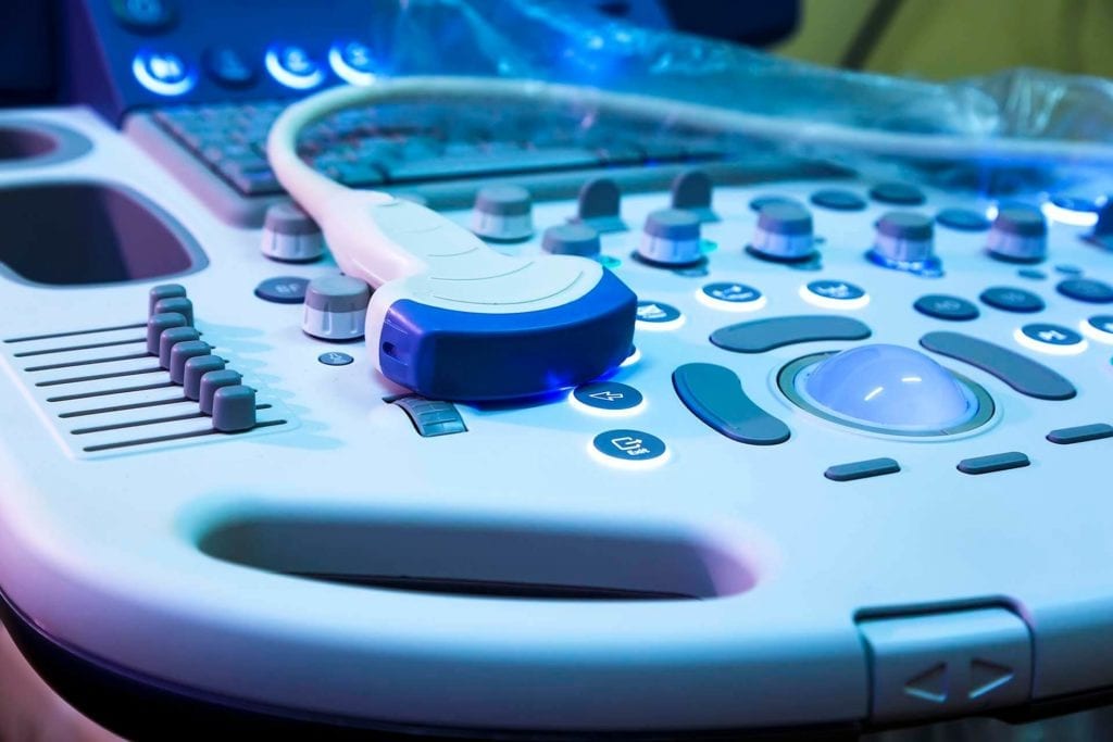
ultrasound at women's imaging center

In addition to breast ultrasound, additional ultrasound exam types are also performed at the Women's Imaging Center. Ultrasound (sonography) utilizes sound waves to produce images of different body parts. Ultrasound exams produce no radiation to patients and are therefore preferred as the initial tests when possible depending on the area being evaluated, especially in young patients and children.

Preparation:

You should wear comfortable, loose-fitting clothing for your ultrasound exam.

Preparation protocols depend on the type of ultrasound you are having. You will be provided with specific instructions when scheduling your exam. Examples include:

  • For a study of the abdomen, patients are asked to avoid eating for 6 to 8 hours before the test.
  • For ultrasound of the kidneys or pelvis, you may be asked to drink four to six glasses of liquid about an hour before the test to fill your bladder.

For additional information about specific tests, please follow the links below:

Abdominal Ultrasound
Carotid Ultrasound
Obstetrical Ultrasound
Pelvic Ultrasound
Scrotum Ultrasound
Thyroid Ultrasound
Vascular Ultrasound
Venous Ultrasound

OB Sonograms

Obstetric sonograms are ultrasound studies that specifically evaluate the baby (fetus) and maternal organs during pregnancy.  Sound waves are used to produce real-time images without ionizing radiation. The test is noninvasive and has no known harmful effects.

Additional information may be found at Radiology.org.

OB Sonogram
WIC_Color_Icon

Schedule your appointment today!